
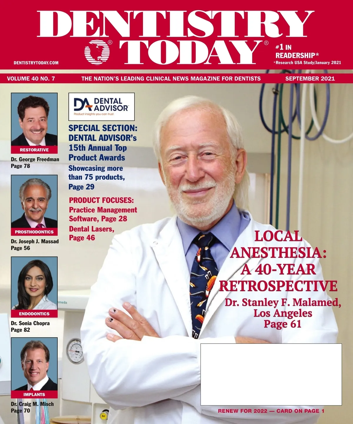
September 2021 · 92 pages
Dentistry Today — September 2021
Clinical articles, techniques, product news and continuing education for dentists. Read the full issue free in the digital edition.
Inside this issue
Excerpts from the issue. Open the digital edition to read any page in full.
- Page 8
The Long and Winding Road As we approach fall, I had great expectations of how this year was going to end. I went to the Florida Dental Association meeting in June and things started looking normal. Meeting planners got in touch with me and suddenly, I was on the booking flights through the end of the year to Utah, California, Maryland, Nevada, Kentucky, and Washington state. Governors were lifting restrictions and I put my stockpile of N95 masks in my garage. Then the Greek alpha bet soup began to appear. As of the writing of this column, several state and…
- Page 10
Post-Pandemic Fraud in Dental Practices INTRODUCTION Prosperident has been in the business of investigating embezzlement committed against dentists since 1989. Over that time, we have seen many changes in both dentistry and the environment in which it operates. If you need help with the word embezzlement, it refers to someone in a position of trust (often a staff member, but also potentially someone external like a bookkeeper) abusing that position and the trust to steal. The administration of practices has evolved greatly in the last 30 years. From the company's initial pegboard investigations when few practices were computerized, to the…
- Page 12
VIEWPOINT continued from page 10 zled. Results showed 5% reported never, 26% said once, 11% said twice, 2% said three times, and 8% said more than three times. While it is too soon to quantize the impact of the pandemic on the prevalence of embezzlement, we know from watching business cycles over many years that booms and busts each pressure a different kind of embezzler. People normally steal for 2 reasons: some embezzle because of perceived need. For whatever reason, these people are unable to meet their monthly financial obligations, and they fall further behind over time until their basic…
- Page 14
Innovative Handling of Soft Tissue Around an Immediately Placed Dental Implant The role of attached keratinized gingiva (AKG) around a healthy dental implant cannot be over emphasized. AKG forms an important seal between the oral cavity and the implant beneath the soft-tissue junction. It also contributes to thickness and robustness of the peri-implant soft tissue which, in turn, contributes to blood flow and immune support in the crucial soft and hard tissue interface on the face. More recent evidence reminds us that the thickness and quality of the peri-implant tissue are much more important factors that influence preservation of crestal…
- Page 16
Ransomware Steve White discusses the No. 1 threat to your patients' data—cyberattacks—and how to tell if your practice is protected. Q: Are dental offices at risk of ransomware? A: Yes, dental offices and all healthcare providers are under cyberattacks, and the rate and sophistication of these attacks are rapidly increasing! Healthcare was the No. 1 industry attacked in 2019. In 2020, the number of successful attacks more than doubled, and 2021 is on pace to again more than double the number of episodes before the year's end. Yet, at this point, the most frequent comment we hear from offices is,…
- Page 18
Pulp Vitality Tester Accommodates All Dentists' Settings Preferences EDGEWOOD, New York--Parkell's Digitest 3 Pulp Vitality Tester electrically stimulates the tooth to test for vitality. Comparing the response to the stimulus of the suspected tooth with a known healthy tooth's response can help dentists hone in on the source of the patient's pain. As quarantine restrictions ease and patients return to the operatory to have long overdue needs addressed, it has never been more important for clinicians to have every diagnostic tool at their disposal. With the ability to offer the additional diagnostic test of electric stimulation, dentists are discovering they…
- Page 20
Curing Light Features Corded, Cordless Models SOUTH JORDAN, Utah–The VALO Grand curing light from Ultradent Products uses a custom, multi-wave length LED for producing high intensity light at 395 to 515 nm through an optimally collimated beam capable of polymerizing all light cured dental materials, including porcelain and underlying cements. The VALO Grand light features a 12-mm lens. Its extremely durable, slim, ergonomic shape allows unprecedented access to restoration sites. For more information, call Ultradent Products at (800) 552-5512 or visit ultradent.com. FREEinfo, sani-treet plus Enzymatic Cleaner Evacuation Cleaner Instrument Pre-Soak Ultra-Sonic Cleaner Non-toxic, Biodegradable Eliminates organic odors Breaks down…
- Page 22
Disposable Bite-Wing/Sensor Positioner Helps Reduce Clinical Set-Up Time FERNANDINA BEACH, Fla.–Steri-Shield's Wingers Integrated Barrier, a disposable bite-wing, features 3 products in 1. Unlike any other sensor positioner on the market today, this unique product provides a sensor holder already inside a padded, elastic, and seamless infection control barrier. This reduces clinical set-up time to obtain a radiological image from approximately 60 seconds to 5 seconds. It produces an average cost-saving time of 20% compared to purchasing 3 separate items. The seamless elastic barrier eliminates pinholes that may occur in the heat-sealed seams of plastic film sensor barriers. It fits all…
- Page 23
Battery-free Portable Generator Guarantees Consistent Production of X-Rays ATLANTA-Carestream Dental's CS 2400P is a portable generator that offers practices a power full x-ray beam needed for exceptional diagnostic images, yet small enough to be handheld. Thanks to its exclusive battery free design, the CS 2400P can go from completely discharged to ready-to-go in less than 3 minutes and delivers multiple shots before needing to be recharged—which only takes seconds—to keep a team on schedule. Its innovative battery-free concept guarantees a consistent production of x-rays over a charge, unlike battery-operated systems. The CS 2400P's exposure parameters are similar to those of…
- Page 24
QUICK TECHNIQUE C2F (Conversion 2 Final): Small Hole Technology Enables an Efficient Extraoral Conversion That Preserves Occlusion Figure 1. Attach the C2F analogs to the RAPID Appliance and seat onto the model. Figure 2. Using light-curable acrylic resin, lute the analogs to the model. Figure 3. Replace the RAPID Appliance with the prosthetic. Using the hollow C2F analog as your guide, drill 2.4-mm holes through the prosthetic at each site. Figure 4. Seat temporary copings and ensure passivity in the prosthetic. Figure 5. Lute the prosthetic to the temp copings and cure. Fill any remaining voids and finish. Figure 6.…
- Page 26
CLINICAL REVIEWS DENTAL ADVISOR ASAP INDIRECT+ ★★★★★1/2 (91%) Clinician's Choice Dental Products | (800) 265-3444 | clinicianschoice.com Consultants' Comments • "Easy to use, efficient polishing, and great results." • "It's easy to reach the surface accurately with these polishers." • "I used it on zirconia crowns, and the luster was great." • "The adjusters were very effective." • "The spiral wheels adapt well to all surfaces and can get into tight areas." • "I like that you can attach it to a latch slow-speed handpiece for intra-oral use." • "Good selection of shapes." • "The polishers left a surface that…
- Page 31
New! Every shade. One choice. omnichROMAFlow One-Shade Flowable Composite OMNICHROMA FLOW is the world's first one-shade flowable composite to esthetically match every color of tooth from A1 to D4 with a single shade of composite. Tokuyama Dental's Smart Chromatic Technology, first implemented through the universal composite OMNICHROMA, has changed the way thousands of dentists practice restorative dentistry forever. Now, the same one-shade benefits are made available in a flowable consistency capable of addressing a wider range of restorative needs, saving clinicians even more time and money. Outstanding Features • Unprecedented shade matching • High polishability • High stain resistance •…
- Page 42
Cordless Endodontic Motor Helps to Avoid File Separation LAS VEGAS–Mididenta's ACE Advanced Cordless Endodontic motor is a compact, continuous, and reciprocating ergonomically designed cordless base, cordless motor with an accurate built in apex locator. It's a silent, brushless motor with a 6:1 mini slim neck contra angle and Proprietary Genius AutoSense technology that helps to avoid file separation. The ACE also features 8 customizable rotary settings and 3 reciprocation and preset file programs for easy use. It offers a lot of auto safety features. For more information, call Mididenta at (800) 221-0750 or visit the website mididentа.com. FREEinfo, Thin Articulating…
- Page 44
Devices Relieve Tension Headaches and Migraines NEWPORT BEACH, Calif.— Glidewell now supplies dentists with 2 devices proven effective at relieving their patients' tension headaches and migraines: the NTI tss Plus and the NTI OmniSplint. By adding this new clinical opportunity to its family of preventive care products, Glidewell has made it easier than ever for dental clinicians to help the 30 million people in the United States who suffer from migraines (data on file). The NTI tss Plus has been prescribed by thousands of dentists for more than 20 years and is a trusted name in the industry. The device…
- Page 46
Dental Lasers DIODE LASERS AMD Lasers Diode lasers can improve ginggivectomies, frenectomies, and more. Because lasers coagulate while they cut, procedures are performed in the most ideal. Precision Lasers are some of the most celebrated, endorsed, and awarded dental lasers. AMD Lasers' mission is to ensure every dental professional has access to advanced technology and education that will elevate the standard of patient care. For more information, call (866) 999-2635 or visit the website located at amdlasers.com. FREEinfo, BIOLASE | Epic Hygiene (833) 246-5273 | biolase.com/epichygiene FREEinfo, CAO Group Precise LTM soft-tissue diode laser facilitates everyday procedures and easily moves…
- Page 48
Industry News Delta Dental of California Named Among Top Bay Area Corporate Philanthropists Delta Dental of California announced it has been recognized as one of the top 100 corporate philanthropists in the San Francisco Bay Area by the San Francisco Business Times. In the publication's 2020 Bay Area Corporate Philanthropists list, Delta Dental is ranked number 27, marking the fourth consecutive year of a placement in the top 50 and reflecting the dental industry's vital role as a benefit provider's continuing commitment to investing in the communities which it serves. "Twenty-twenty was a challenging year for everyone and especially our…
- Page 52
Adhesives: An Invaluable Asset for Managing Denture Patients Joseph J. Massad, DDS Lily T. Garcia, DDS, MDS Swati Ahuja, BDS, MDS For a complete denture to be successful, it should be retentive, stable, and optimally supported by the denture-bearing tissues.1 Despite following the best prosthodontic practices, the presence of certain patient conditions, such as poor quantity and quality of saliva, extensive loss of volume of hard and soft tissues, and/or neuromuscular disorders may negatively affect the retention and stability of complete dentures.2-6 A prosthesis that dislodges at rest or during functional movements may lead to dysfunctional oral activities, tissue trauma,…
- Page 57
powered (electric) toothbrushes when compared to manual toothbrushes.22-23 Electric toothbrushes are preferred by most patients because they are efficient, easy, and convenient to use.24 Recently, newer electric brushes (Genius [Oral-B]) with 3D cleaning action technology (oscillation, rotation, and pulsation), pressure sensors, position detection, and built-in timers have been introduced. The oscillating, rotating, and pulsating action of the electric toothbrush stimulates and massages the tissues, thereby increasing the velocity of red blood cells, which carry oxygen into the tissues. The use of electric toothbrushes for preventing peri-implantitis and maintaining peri-implant health has been discussed in several articles.14-18 CLEANING THE ORAL CAVITY…
- Page 58
Figure 4a. The Genius positioned on the palatal/lingual surfaces of the natural teeth. Figure 4b. The brush head positioned on the occlusal/incisal surfaces of a fixed dental prosthesis. Figure 4c. The brush head positioned at the junction of the implant-supported fixed complete denture and the tissues. Figure 5a. The Genius interproximal brush head positioned for use in interdental cleaning. Figure 5b. The interproximal brush head positioned over the pontic and the tissues. Figure 5c. The interproximal brush head positioned between the prosthesis and the tissues. Denture adhesive use is common among denture wearers. It is important to teach patients an…
- Page 61
Dental Pain Control and Local Anesthesia: A 40-Year Journey INTRODUCTION Local anesthetics (LAs) form the backbone of pain control techniques in dentistry. Modern dentistry began with the introduction of cocaine in 1885 and procaine (Novocain) in 1905, allowing dentists and surgeons to painlessly carry out procedures that previously had been impossible to do or were excruciatingly painful. From the early 1900s until the mid-1940s, the "ester-type" LAs were used: Procaine, propoxycaine, and tetracaine were the most popular. In 1948, the first "amide-type" LA, lidocaine (Xylocaine), was introduced, quickly becoming the gold standard. The 1960s and 1970s saw the introduction of…
- Page 62
ANESTHESIA [Figure 1 shows chemical structures of three molecules:] Procaine - Structure with H2N group and ester bond Lidocaine - Structure with CH3 groups and amide bond Articaine - Structure with COOCH3 group and amide bond [Legend shows: Ester bond (blue), Amide bond (dark red)] Figure 1. Chemical structures of an ester (procaine), amide (lidocaine), and articaine. [Figure 2 shows a line graph with time (minutes) on x-axis (0-90) and C (mg/l) on y-axis (0-1.2). Two lines are plotted showing plasma levels of lidocaine and articaine over time, with both declining after reaching peaks around 10-15 minutes.] Figure 2. Plasma…
- Page 64
Dental Pain Control and Local... continued from page 62 There are 2 distinct pharmacokinetic and chemical characteristics that make articaine popular:(1) more rapid elimination from the cardiovascular system (elimination half-life) and (2) lipid solubility. More rapid elimination leads to increased safety, and it's being a preferential drug, in this author's opinion, in patients who are pregnant, nursing, geriatric, and pediatric, while being more lipid-soluble leads to more rapid onset and greater depth of anesthesia. Additionally, articaine's greater lipid solubility enables it to be successful using injection techniques that have been uniformly unsuccessful with other LAs. Elimination half-life: Classified as an…
- Page 65
ANESTHESIA "Modern" dentistry began with the introduction of cocaine in 1885 and procaine (Novocain) in 1905, allowing dentists and surgeons to painlessly carry out procedures.... anesthesia is frequently necessary for dental procedures. Clinical trials comparing the efficacy of articaine and lidocaine in providing palatal soft tissue anesthesia following prelabiary bi level dentistrais stratied articaine's superiority. Gholami et al11 repeated the infiltration of 0.6 mL of LA at 2-minute intervals and evaluated palatal anesthesia. At 4 minutes (1.2 mL), 65.4% of articaine patients and 0.65% of lidocaine patients had palatal anesthesia. At 6 minutes (1.8 mL), values were 82.7% vs 1.3%.…
- Page 67
SPECIAL ADVERTISING SECTION 67 CASE STUDY A Clinician's Positive Results Using the Ortek-ECD A 22-year-old patient came to the office complaining of tooth sensitivity when eating sweets. Clinical examination showed obvious decay in the disto-occlusal pit on the maxillary first molar (Figure 1, site 3). Upon further examination, the mesio-occlusal pit (Figure 1, site 2) was suspicious, but there was no evidence of cavitation from visual and radiographic methods, making a treatment decision difficult. A second mesio-occlusal pit appeared sound (Figure 1, site 1). This case was ideal to evaluate the digital caries score indicated by the Ortek-ECD, as there…
- Page 68
Setting the Foundation for Healthy Implants Timothy F. Kosinski, DDS Setting the foundation for ideal dental implant placement requires precise preparation of the hard tissue, an understanding of hard tissue contours and vital anatomy. Just like building a house, a solid foundation ensures that a beautiful home is created and remains stable for many years. Shortcuts in this process results in improperly chosen implant size or shape which may result in overstressing of the bone around the fixture and eventual breakdown or loss. Making sure our patients clearly understand the value of crafting socket sites following non-restorable tooth removal is…
- Page 70
Guided Bone Regeneration: Membrane Selection INTRODUCTION Guided bone regeneration (GBR) is the most common method used for bone augmentation in implant dentistry. GBR utilizes a barrier membrane to occlude soft tissue cells and allow slower growing bone cells to repopulate the defect and regenerate bone. Additional principles for successful GBR include tension free primary closure of the flap over the site, angiogenesis during healing to provide the necessary blood supply and undifferentiated mesenchymal cells, space creation, and maintenance for bone ingrowth, and stability of the wound during healing. The type of membrane used for GBR is based on several factors,…
- Page 72
IMPLANTS FIGURE 1. Preoperative view of 2 failing dental implants replacing the mandibular incisors. FIGURE 2. A periapical radiograph reveals significant bone loss around the implants and adjacent teeth. FIGURE 3. The healed ridge after implant removal. FIGURE 4. The ridge is exposed for cortical perforations and harvesting autogenous bone. FIGURE 5. Tenting screws were placed in the anterior mandible to support the graft. FIGURE 6. The ridge was augmented with particulate autograft, bovine bone mineral, and platelet-rich fibrin. FIGURE 7. The collagen membrane is secured with periosteal sutures. FIGURE 8. The reconstructed ridge after 6 months of healing. FIGURE…
- Page 73
IMPLANTS 73 Figure 14. Pre-op view of the failed maxillary implants supporting a fixed bridge. Figure 15. Periapical radiograph of the maxillary left implant reveals severe marginal bone loss. Figure 16. The maxillary ridge following healing after implant removal. Figure 17. Exposure of the atrophic maxilla. Figure 18. A tenting screw is used to support the particulate bone graft and membrane. Figure 19. The ridge was augmented with particulate autograft, bovine bone mineral, and platelet-rich fibrin. Figure 20. The graft in the left maxilla was covered with collagen membrane and secured with periosteal sutures. Figure 21. The graft in the…
- Page 74
Using Alloplastic Material for Subantral Grafting INTRODUCTION When maxillary posterior teeth are extracted, and the extraction site is not grafted, the maxillary sinus can pneumatize. Even in areas where a graft was placed but an extended period of time has passed, this enlargement of the sinus cavity, along with the loss of crestal bone volume, can complicate the placement of dental implants. The complication revolves around the loss of height of bone that prevents implant placement due to not enough vertical height to contain the implant without perforating into the sinus or not having enough bone to resist prosthetic loads…
- Page 75
Figure 1. OsteoGen Plug (IMPLADENT LTD). Figure 2. The vertical, or Summers, approach. Figure 3. Reflected soft tissue on ridge. Figure 4. Placement of a 4.3-mm-diameter paralleling pin. Figure 5. A radiograph to check the depth and position of the osteotomy. Figure 6. Concave osteotomes. Figure 7. OsteoGen Plug. Figure 8. The plug, adapted for placement in the osteotomy. Figure 9. Torquing the implant into position. No. 15 blade and a periosteal elevator exposed the crestal bone. I prefer to do full thickness elevations when doing crestal lifts so I can see the crest and have a definitive reference point…
- Page 76
Figure 10. Penguin RFA. Figure 11. Sutured surgical site. Figure 12. Immediate postoperative radiograph. Figure 13. A 3D image of the graft after healing. Figure 14. Lateral wall approach. Figure 15. Preoperative pneumatized sinus. Figure 16. Outline of the lateral window. Figure 17. Sinus curettes. Figure 18. OsteoGen Strips (IMPLADENT LTD) in saline. Figure 19. Strips placed in the elevated sinus. Figure 20. Implant placement. Figure 21. Filled sinus cavity. Figure 22. Collagen membrane in place. Figure 23. Surgical site closed with sutures. Figure 24. Immediate post-surgical radiograph. Figure 25. A 4-month radiograph of the augmented site and implant. shows…
- Page 77
IMPLANTS conform to the contours of the sinus cavity. The OsteoGen Strip has the same makeup as the OsteoGen Plugs but in a strip form that can be used for grafting around implants in immediate placement cases and a host of other grafting scenarios. Starting at the most posterior aspect of the initial elevation, the soaked strips were placed and gently compacted against the walls (Figure 19). Working from posterior to medial and then to anterior aspects of the lift, additional strips were placed to fill any voids. A 5.0 × 10-mm Hahn Tapered Implant System was placed with 20…
- Page 78
Restorative Aesthetics at the Gingiva AESTHETIC DILEMMA OF THE GINGIVAL MARGIN The data is clear: Phenolic and composite resin are both biocompatible at the gingival margin and well-tolerated.1-3 The major cause for concern is that composite can cause tissue irritation if it impinges directly on the gingiva.4-8 This may ultimately cause rather unaesthetic gingival recession. Porcelain is less challenging, but marginal plague buildup, often stimulated by the thin layer of exposed resin cement, can lead to a similar irritation of the free gingiva and, ultimately, recession.9-12 Even skilful subgingival margin placement will typically, within 3 to 5 years, whether due…
- Page 80
Restorative Aesthetics at the Gingiva continued from page 78 Hence, Giomer fluoride releasing capacity does not decrease over time. It is essential that the restorative gingival margin (whether pink or enamel in color) is positioned supragingivallyand somewhat away from the free gingival margin.3,4,5 While a very narrow darker band of dentinal structure may be visible apically, the restoration's coronal enamel and pink gingival coloration focus attention away from this region. Gingivally blended restorations allow the professional to deliver both aesthetic and supragingival margins in the same restoration. The supragingival margin also facilitates effective home maintenance for the patient. Restorations that…
- Page 82
The Endodontic Renaissance and Modern-Day Root Canals INTRODUCTION Endodontics has come a long way! I remember when I became an endodontic resident, many of the dental school instructors told me I was entering into a dying specialty. Since then, so many new technologies have come to the scene to give my specialty new life. I am excited about this new revival. There is no doubt that tooth anatomy is a huge obstacle when it comes to disinfecting teeth. Tooth anatomy is probably the No. 1 culprit for failed root canals, and yet, it doesn't get enough attention. When most dentists…
- Page 83
CHAIRSIDEWITHDRC.K THE SECRET TO MORE EFFECTIVE WHITENING No two patients are exactly the same. So why use just one whitening protocol to treat them all? At KoR Whitening, we recognize that each patient presents a different whitening challenge and that's why we developed a full line of whitening systems to meet your patient's individual need. And KoR Whitening is the first whitening system in the world to maintain potency of its entire line of whitening gels by constant refrigeration from the instant of manufacture until being received cold-packed at your office. Dr. Rod Kurthy #1 Proper Product and Protocol KoR…
- Page 84
ENDODONTICS CASE 1. Figure 1. Straight-angle PA. Figure 2. Off-angle PA. Figure 3. Bite-wing. Figure 4. Intraoral clinical photo. Figure 5. Straight angle postoperative radiograph. Figure 6. Off angle post op radiograph. Figure 7. Axial slice CBCT scan. Figure 8. Coronal slice CBCT scan of the coronal third of the canal. Figure 9. Coronal slice CBCT scan of the apical third of the canal. CASE 2. Figure 10. Straight-angle preoperative radiograph. Figure 11. Off angle pre-op radiograph. Figure 12. Bite-wing radiograph. Figure 13. Sagittal view CBCT scan. Figure 14. Coronal view CBCT scan. Figure 15. Axial view CBCT scan. The…
- Page 85
ENDODONTICS 85 CASE 3. Figure 16. Straight-angle pre-op radiograph. Figure 17. Off-angle pre-op radiograph. Figure 18. Bite-wing radiograph. Figure 19. Conefit radiograph. Figure 20. Off-angle post-op radiograph. Figure 21. Straight-angle post-op radiograph. Figure 22. Pre-op radiograph No. 30. Figure 23. Pre-op sagittal view CBCT scan. Case 4 In examining one more case, the anatomy, once again, was something I wasn't expecting. In the pre-op radio graphs, notice the large periapical radiolucency on the mesial root (Figures 24 to 26). The treatment looked pretty straightforward, and the procedure seemed to be going along predictably. Once again, the GentleWave was used on…