
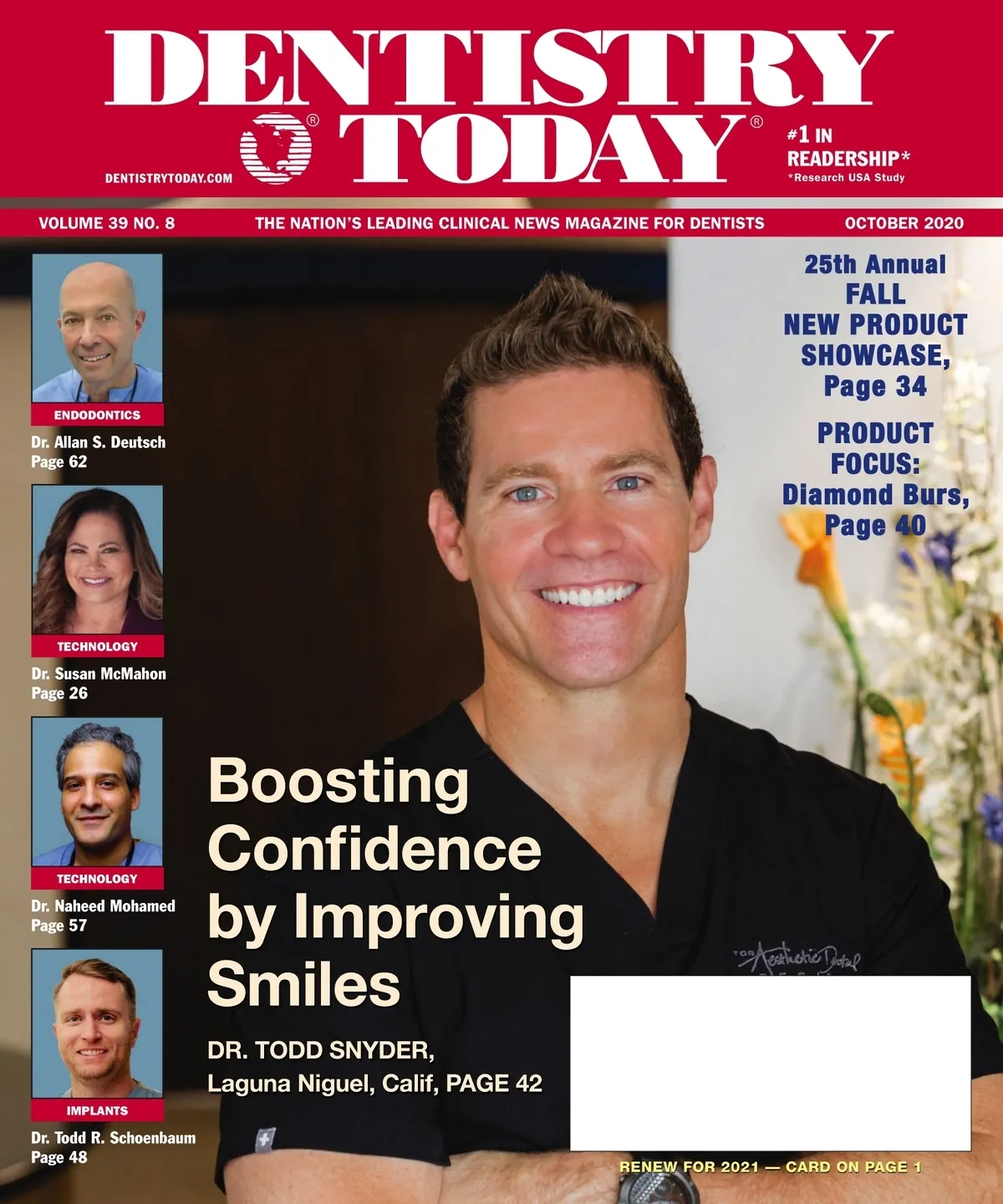
September 2020 · 72 pages
Dentistry Today — October 2020
Clinical articles, techniques, product news and continuing education for dentists. Read the full issue free in the digital edition.
Inside this issue
Excerpts from the issue. Open the digital edition to read any page in full.
- Page 8
Missing the In-Person Dental Meetings? As we arrived at the first full month of fall, it seemed strange, to say the least, that we were not preparing to physically attend the annual meeting of the American Dental Association! Since all the meetings for the remainder of 2020, and some now in early 2021, have been rightly and responsibly converted to virtual meetings due to the COVID-19 pandemic, it will be interesting to see how success will be measured these events. What will the post-event evaluations look like in terms of the level of involvement/attendance as well as the post-meeting evaluations…
- Page 10
The Courage to Be Vulnerable Together By the time I turned 40, I assumed that I would have had life all figured out. More importantly, I thought that I'd have depression figured out. Because even to this day, despite what I have read, been taught, or been told by my treatment team, I still look at depression as a sign of my own weakness. Within my soul, I feel like it is something I should have control over. And contrary to what I believe, this disease isn't a weakness. However, just like many other people who are plagued by depression,…
- Page 12
VIEWPOINT continued from page 10 nity. Acknowledging and admitting this common problem will help in dispelling shame and the falsity that depression is a weakness. Sharing our stories and relating to one another will lighten the burden when examining our own self-perceived negative identities. We all have to wear a mask in front of our patients, our team members, and sometimes even our families; let's not wear that mask in front of one another. In college, I suffered a significant setback on account of my depression. I took some time off. Following my hiatus, I walked back into the university…
- Page 14
Look Behind the Scenes Marty Jablow, DMD, focuses on the mission and selection process for the yearly Cellerant Best of Class Technology Awards. Q: What are Cellerant Best of Class Technology Awards about? A: The 12th annual Cellerant Best of Class Technology Award winners have been selected. This award is not a popularity contest but an exercise that is focused on thoughtfully selecting new and established products and technologies that can have a very positive impact for your dental practice. Q: Can you please explain what is involved in the selection process? A: Following a year of research, the selection…
- Page 16
CELLERANT BEST OF CLASS TECHNOLOGY AWARDS 2020 Back in February, the committee for the Cellerant Best of Class Technology Awards met for the 12th year at the Chicago Dental Society's 155th Midwinter Meeting and voted on the Best of Class products of 2019 and a few that emerged in the first few months of 2020. As in past years, there were several products that were previous winners and a few new innovations that had not been seen in dentistry before. The committee members talk with manufacturers, scour dental meetings throughout the year, and sit through detailed presentations. Many of the…
- Page 18
From the first phone call to the final payment. Weave's unique integration of hardware and software solutions help businesses grow, retain and communicate across the entire customer journey. Schedule a live demo today. 888.545.8880 getweave.com/dentistrytoday WEAVE PHONES WEAVE MESSAGES 7 SYSTEMS IN 1 WEAVE CORE WEAVE CUSTOMER INSIGHT WEAVE SCHEDULING WEAVE PHONES WEAVE TEAM WEAVE PAYMENTS WEAVE ANALYTICS weave CELLERANT BEST OF CLASS 2020 FREEinfo, on card --- BEST OF CLASS TECHNOLOGY AWARDS Weave Weave | (888) 579-5668 | getweave.com Communication with our patients is a primary foundation of any dental practice. Especially in this current, ever-changing time, the office…
- Page 19
TeleDent MouthWatch | (877) 544-4342 | mouthwatch.com MouthWatch has been a pioneer in the teledentistry space for many years. Its TeleDent product allows dentists to easily communicate via web and mobile-friendly messaging and real-time video with patients, colleagues, specialists, and others using a secure, HIPAA-compliant portal. Although we made this choice in February, it has become one of the most relevant systems we've used in this new, unique time. MouthWatch founder and CEO Brant Herman has pushed the process to new levels, making it efficient for practitioners to keep in touch with everyone not only in crisis situations but also…
- Page 20
Why Teledentistry Is Here to Stay Riddhi Gangolli, BDS, MS, PhD, explains the value of integrating telehealth into your practice. Riddhi Gangolli, BDS, MS, PhD Q. The pandemic has served as a turning point for telehealth. How can dental practices benefit from teledentistry? A. Teledentistry, including SmileDirectClub's clear aligner therapy treatment, but we created the Partner Network in early 2020 to open our teledentistry platform to more dentists and orthodontists. This gives them the opportunity to expand their current services and provide patients with a truly get started at a SmileDirectClub-affiliated dentist's office if they choose. The SmileDirectClub Partner Network…
- Page 22
Sanitizing Station Helps Stop Spread of Germs NEWBERG, Ore—Now that dental practices have reopened their doors, patients are more concerned than ever about what's being done to prevent the spread of germs. The A dec Protect and Sanitize Station provides peace of mind—and a healthier waiting room area—by offering patients access to hand sanitizer, tissues, and face masks on one convenient stand. The unit, which needs only minor assembly, includes a touchless hand sanitizer dispenser with a drip shield and one Purell cartridge. It's also available to order without the Purell dispenser for clinicians who prefer a different brand. An…
- Page 24
Suite of Technologies Allows for Completing All Components of Restorations in One Appointment Whitening System Designed for Treating Tetracycline IRVINE, Calif.- The KoR Ultra-T whitening system is designed specifically to treat tetracycline and has even proven effective on dentinogenesis imperfecta cases. The system utilizes an in-office "turbo" conditioning application prior to at-home whitening, using KoR Dual-Activated, Tri-Barrel 13% Hydremide Peroxide. This gel is placed into KoR Seal Trays, and no retractors or paint-on gingival barriers are necessary. Two consecutive 20-minute applications are required. The 13% Hydremide Peroxide is specifically catalyzed in a way to initiate the internal cleansing of the…
- Page 25
Dentists Doing Botox? Now More Than Ever! The phrase "Botox in dentistry" has become synonymous with the American Academy of Facial Esthetics, better known as the AAFE. Founded in 2008 by internationally known dental educator and clinician Dr. Louis Malmacher, the AAFE has quite literally set the standard for live patient certification education for Botox, dermal fillers, and solid-filler PDO thread therapy in dentistry and medicine. "When we started the AAFE, Botox use was almost unheard of in dentistry," Malmacher said. "We recognized the tremendous potential that Botox and dermal fillers had in every dental discipline, including restorative implants, periodontics,…
- Page 26
The Next Dimension Why General Practitioners Need a CBCT Machine Cone beam computed tomography (CBCT) has motivated a new standard of care when it comes to diagnosis of dental diseases by way of 3-D imaging of the maxillofacial skeleton. For nearly 2 decades, CBCT technology has been available, but only recently has it been commercialized and popularized in the field of digital dentistry. Two dimensional diagnostic imaging, such as traditional radiographs, photographs, and video imaging, are essential to dentistry, no doubt, but limited. Magnification, geometric distortion, and projective displacements may all be misconstrued with a 2-D imaging. In contrast, 3-D…
- Page 28
Figure 2. A CBCT scan was taken to assist in the diagnosis of symptoms on the patient's mandibular right side. See lucency at the lower right first and second molar not evident on the 2-D radiograph. Figure 3. Axial view of the root apices of the mandible right first molar on the CBCT scan. The Next Dimension: Why General... continued from page 26 this patient to an endodontist for evaluation, but there's no telling if the patient will go to the referral or wait until the problem is exacerbated. In having a CBCT machine in-office, a full diagnosis is available…
- Page 30
New Retractor Comfortably Keeps Mouth Open, Avoids Muscle Straining SOUTH JORDAN, Utah–The new Umbrella lips, cheek, and tongue retractor from Ultradent Products has been specially designed and tested for 2 simple purposes: patient comfort and quick, easy placement. Keeping the balance of a stable retractor that gives the patient a serene, relaxing experience is no major task. It took years of research and development to get the precise dimensions and architecture to be anatomically correct. This retractor keeps the patient's mouth open in the 2- to 3-mm range so that it's comfortable and doesn't strain the muscles. The name Umbrella…
- Page 31
CHAIRSIDE WITH DR.K THE SECRET TO MORE EFFECTIVE WHITENING No two patients are exactly the same. So why use just one whitening protocol to treat them all? At KöR Whitening, we recognize that each patient presents a different whitening challenge and that's why we developed a full line of whitening systems to meet your patient's individual need. And KöR Whitening is the first whitening system in the world to maintain potency of its entire line of whitening gels by constant refrigeration from the instant of manufacture until being received cold-packed at your office. Dr. Rod Kurthy #1 Proper Product and…
- Page 33
INNOVATIONS 33 Bioceramic Releases Calcium Ions, Helps Form Mineralized Tissue LONDRINA, Parana, Brazil–BIO C SEALER ION+ from Angelus USA is a resin free bioceramic that releases calcium ions that help in the formation of mineralized tissue. The alkaline pH and excellent marginal sealing offer safety and high rates of success in endodontic treatment. In addition, the premixed, ready-to-use formulation effectively simplifies the obturation procedure, results in less chair time and more patient comfort. For more information, call Angelus USA at (855) 346-3682 or visit angelusdental.com. FREEinfo, SPOTLIGHT Flip-Up Face Shield Provides Ability to Mount Any Loupes or Eyewear CENTENNIAL, Colo.–The…
- Page 34
DENTISTRY TODAY 25th ANNUAL FALL NEW PRODUCT SHOWCASE Dentistry Today's 2020 Fall New Product Showcase highlights the latest products being offered to dental professionals. The products appear in no particular order. exocad America SINGLE-VISIT DENTISTRY SOLUTION The ChairsideCAD 2.3 Matera has the widest range of indications for single-visit dentistry and the broadest selection of integrated open architecture devices. The software is intuitive and easy to learn, so users reliably deliver esthetically demanding restorations in a single patient session. ChairsideCAD and modules are integrated using exocad's proven and robust software platform successfully in use for more than 10 years. For more…
- Page 35
From the innovators at Angelus comes ONE SINGLE POST WRAPPED IN ONE SIMPLE SOLUTION INTRODUCING SPLENDOR-SAP SINGLE ADJUSTABLE POST Universal Anatomical Highly retentive Minimally invasive Low risk of root fracture angelus A universal system using a single drill, a single sleeve, and a single post to create a single solution for restoring a compromised tooth. Reduces the number of drills and posts needed and increases post adaptation in the cervical region. Universal Post Optional Sleeve Optimized Fit angelusdental.com | 1.855.346.3682 Patent Pending. Available in the U.S. FREEinfo, on card Available through HENRY SCHEIN DENTAL (1-800-372-4346) KöR Whitening WHITENING SYSTEMS KöR…
- Page 36
Dental Herb Company TREATMENT GEL PerioSpot is a powerful treatment gel to soothe and reduce harmful bacteria and gum inflammation. Apply this concentrated gel post-procedure to jumpstart the healing after SRPs, implants, crown preps, etc. It is ideal for patient home care to provide continued relief and protection. PerioSpot also relieves stubborn inflammation and mouth sores. For more information, call (800) 747-4372 or visit dentalherb.com or periospotel.com. FREEinfo, Align Technology IMAGING SYSTEM The iTero Element 5D imaging system is the first powerful hybrid platform that optimizes workflows by simultaneously recording 3-D images, intraoral color, and near-infrared imaging (NIRI). NIRI aids…
- Page 38
SmileDirectClub ELECTRIC TOOTHBRUSH This electric toothbrush is a powerful, premium, oral toothbrush that cleans 50% better than a manual toothbrush, thanks to quiet, sonic vibration that moves bristles, not the handle. A 2-minute timer with quadrant timing ensures even brushing in the right areas for the right amount of time. The smart design is roll-resistant, ensuring the electric toothbrush stays put, and includes a versatile 3-in-1 case. For more information, visit smiledirectclub.com. FREEinfo, Microcopy DIAMOND BUR Thirty years ago, Microcopy changed the industry when it introduced the single-patient-use Monodimond, which quickly became the industry standard. Not only did the company…
- Page 39
Enzyme Industries ENZYMATIC CLEANER Sani-Soak Ultra is a dual-purpose, anti-corrosive, enzymatic cleaner that can be used in the ultrasonic and as a wet evacuation cleaner. It is highly concentrated, non-toxic, biodegradable, safe for the environment, and effective in removing organic debris while improving the life of instruments and burs. Sani-Soak Ultra is offered in 2 scents (Lemongrass Lavender and Cool Mint) and dispensed in a liquid-pour measured quart or gallon with a pump. For more information, call (888) 236-9963 or visit enzymeindudstries.com/products/sani-soak-ultra. FREEinfo, Filhol Dental PIN FILPIN for dentin retention pulpal pin requirements provides maximum advantages without compromising safety, dentin…
- Page 40
Diamond Burs Microcopy More than 25 years ago, Microcopy changed the industry when the single-patient-use NeoDiamond was introduced. These award-winning diamond burs quickly became the industry standard. NeoDiamond offers Triton bonding and 20% more diamond cutting surface for a faster cut with less chair time. The burs are individually packaged, sterilized, and color coded with Quick Grit ID for safety and convenience. For more information, call toll-free at (800) 235-1863 or visit neodiamond.com. FREEinfo, A&M Instruments The Diamond Burs are crafted from high-quality stainless steel that resists corrosion during sterilization and bonds with a double layer of diamonds during the…
- Page 41
Shofu Dental Shofu Robot Diamonds, one-piece steel shank diamonds, meet strict ISO manufacturing standards. Recognized by independent testing for their superior performance, cutting efficiency, and lack of clogging, Robot Diamonds are made with a multi-coating technique with a special surface treatment for durability. The smooth cutting and long-lasting Robot Diamonds are available in a wide range of grits (regular, coarse, super coarse, fine, and superfine), shapes, and sizes, including a minimally invasive series. For more information, call (800) 827-4638 or visit shofu.com. FREEinfo, Brasseler USA With one of the most extensive diamond instrument lines in the industry, Brasseler USA diamonds…
- Page 42
Dentistry Can Change Lives The impact of a smile often has untold ramifications on a patient. A new smile can often be the catalyst to spark a change in a person's brain chemistry and mindset to put them on a new path in life. A smile being altered can create a chain reaction of events, starting from happiness and excitement, that releases endorphins, serotonin, dopamine, and oxytocin in the brain. This chemical release can be the needed stimuli to reduce cortisol levels in the brain, allowing a person to be happier and more confident, potentially creating a new path and…
- Page 44
Figure 17. Custom healing abutments fabricated from a pick-up impression during the placement of the implants. Figure 18. Custom impression abutments fabricated on the same models as the custom healing abutments. Figure 19. Impression of the maxillary custom impression abutments using thermoplastic trays (HeatWave) and a quality VPS impression material (Affinity [Clinician's Choice Dental Products]). Figure 20. Maxillary arch photo showing the implant restorations with the GOLFU ForteU (GC America) and Velum materials that were placed in the screw-access holes. to be stable with no visible slide being perceived. A hinge axis recording (SAM 3 Articulator) was done to determine…
- Page 45
AESTHETICS made. The beautiful restorations can be seen in Figures 6 and 16. The patient was very pleased with the results thus far. Implants, Restorations, and a Nightmare Next, the dental implants placed by a periodontist, and they were allowed to heal for a few weeks. The implants were uncovered, and custom healing abutments were placed to allow for tissue conditioning and to achieve the desired contour and health over a period of weeks. Next, custom implant impression analogs that mirrored the same shape as the custom healing abutments were used to capture the position of the implants in relation…
- Page 48
Compromised Implant An Interdisciplinary Rescue Treatment INTRODUCTION Implant treatment for a single missing tooth in the aesthetic zone is considered advanced treatment due to its significantly increased complexity. The "rescue" treatment of an implant involves the correction of a previous implant procedure that has resulted in a subpar outcome. In addition to the biological considerations needed for predictable osseointegration,1 the aesthetic zone presents additional challenges of limited interproximal and inter-occlusal space, non axial loading forces and guidance pathways, and aesthetic challenges of replicating the tooth and gingiva.2 Utmost respect and consideration must be given to biologic, functional, and aesthetic factors.3,4…
- Page 50
Figure 1. Preoperative view of the patient at full smile; the photo was from the previous treating clinician. Note the full gingival display. She underwent treatment of tooth No. 9 with an implant at the initial provider. Figure 2. Close-up view of the pre-operative state of tooth No. 9 and the adjacent teeth. Note the normal gingival phenotype and moderate deficiency of the papilla between teeth Nos. 9 and 10. Figure 3. Patient presentation after healing following immediate placement of an implant at the No. 9 position. Note the reasonable volume of the papilla, though blunted. Tissues appear healthy. We…
- Page 51
IMPLANTS uration, the patient was seen for an evaluation. Collectively, it was determined that further surgeries were unlikely to improve the outcome and that the biologic prognosis of the site was good. As initially discussed with the patient, correction of the gingival defect would have to be accomplished with pink porcelain. A balance must be struck between hygiene accessibility and aesthetics. lain. An intraoral scan (TRIOS) was performed using a Nobel ICBR specific scan body. The patient visited the technician at the dental lab for shade selection of the teeth and soft tissues. A screw retained zirconia restoration (Katana Zirconia…
- Page 52
The Emergency Implant INTRODUCTION Implant dentistry has come a long way in helping the general dentist plan and treat the concerns and needs of edentulous patients. Each patient has unique situations that need to be thoroughly evaluated, and, therefore, case appropriate diagnoses and treatment plans are imperative to realize successful outcomes of care. Anatomic limitations should be addressed, as well as surgical considerations and the final prosthetic design. Soft tissue anatomy and implant osseointegration must be understood prior to any surgical intervention, and the clinician must visualize the outcome of a case before beginning it. If there are contraindications or…
- Page 54
The Emergency Implant continued from page 52 Figure 1. The patient presented with a fistula between the mandibular central incisors. Figure 2. A conventional 2-D digital periapical radiograph appeared to show adequate bone, but a buccal defect had developed (tooth No. 24). Root canal therapy was discussed. Figure 3. An analysis of the sagittal view of the CBCT scan indicated a loss of facial plate of bone. Discussion was undertaken as to the long-term prognosis of endodontic therapy vs removal of the tooth and replacement with a dental implant. Figures 4 and 5. The tooth was carefully removed, and the…
- Page 55
Figure 18. Allograft material was placed into the defect area and covered with the resorbable membrane. Figure 19. The reflected tissue was then repositioned and sutured to place. Figure 20. The patient's natural tooth (clinical crown) was used as his interim provisional while awaiting osseointegration. Figure 21. Adjacent teeth were etched, and the natural tooth's clinical crown was bonded into place with no contacts in all excursive movements. Figure 22. Final radiographic confirmation. Figure 23. Following integration of the implant, the attached gingiva was visualized. The implant was ready to be exposed and for an impression to be taken. Figure…
- Page 56
Dental implants have become mainstream and an excellent treatment modality for the replacement of one to several missing teeth. reflected to expose the entire defect, and the membrane is placed at least 2.0 mm apical and mesial and distal to the defective site. This should also be followed on the lingual side. CLOSING COMMENTS endodontic therapy would likely be poor in this circumstance. It was decided with the patient that an appropriate treatment plan included removal of the tooth, immediate implant placement, and grafting of the site to reform the facial plate. Figures 4 and 5 illustrate how the infected…
- Page 57
Custom 3-D Bone Allograft Block Fabrication INTRODUCTION Digital technology has drastically improved all areas of modern medicine and surely will continue to do so. It favors more accurate treatment planning, minimizes patient chairside time, and leads to greater surgical accuracy in modern implant dentistry. More than 10 years have now passed since the technology was commercialized, and it remains one of the fastest growing industries in dentistry with digital scanning, planning, and even printing being widespread in many dental offices. In implant dentistry specifically, many implants require additional bone grafting prior to implant placement to augment lost or missing bone.…
- Page 58
Figure 1. Preoperative image of the edentulous ridge. Figure 2. A 3D model of the pre-op ridge and planned implant sites using CBCT software. Figure 3. A digitally modelled bone graft over the ridge. Figure 4. A custom 3D rendered digital bone graft. Figure 5. Digital orientation of the digital bone mold with the bone graft. Figure 6. CBCT DICOM cross sectional view, showing the digital planning of the guides, molds, implant, and bone graft (shown in yellow). Figure 7. Bone mold orientation for seating is shown with the 3D printed models. Figure 8. The 3D printed bone mold in…
- Page 59
Figure 13. The custom-made 3-D bone graft after 15 minutes. Note the stability of the graft. Figure 14. Elevated flap, exposing the pre-op ridge. Note the narrow ridge requiring a horizontal bone augmentation. Figure 15. Pilot osteotomies drilled with a surgical guide. Figure 16. Osteotomies and placement of Strumann Bone Level Tapered Implants placed with attachments. Figure 17. The custom, patient-specific bone graft, shown here removed from the mold. Figure 18. Occlusal view of the custom bone graft in place as planned digitally. Figure 19. Buccal view of the custom bone graft in the planned position. Figure 20. PRF membranes…
- Page 60
TECHNOLOGY particular grafts with graft stability, owing to the improvements related to PRF. This was based on previous work conducted by Kawase et al that showed that heat treatment with PRF resulted in its improved stability and also extended resorption properties. In the present case, this concept was ...digital printing has become more routine and widespread than ever before.... utilized to create a custom 3-D bone graft with excellent mechanical handling. Simply, one of the bone molds was preplanned and created, the particulate allograft was inserted, and the liquid PRF was inserted. Based on the work by Kawase et al,…
- Page 62
Overcoming Endodontic Procedural Failures: Part 2 (Editor's Note: This article is the second in a series and continues the discussion on this topic. The first in the series was published in November 2019 in Dentistry Today.) Anatomic Considerations As soon as we pick up the handpiece, we are faced with a procedural difficulty: How deep do we drill to get into the pulp chamber in order to find the canals? For at least half of my career, I was under the assumption that the depth of the pulp chamber had to be different for each tooth in every different patient.…
- Page 63
multiple-visit treatments. I only do multiple visit root canal treatment if I cannot find the canals or if I simply do not have the time in my schedule to complete the endodontic treatment. Instrumentation It is interesting to note the general emphasis placed on instrumentation. The reality is that instrumentation only cleans 35% to 50% of the canals, no matter what system or technique is used.18,19 The remaining debris removal and bacterial reduction is accomplished through the irrigation regime. So if all endodontic instrumentation systems clean equally well, what are we looking for in using a particular type of instrument?…
- Page 64
Figure 1. L represents the distance between the tip of the bar and where the conical shaft meets the cylindrical shaft. That distance is approximately 6.50 mm. L = 6.50 mm Figure 2. The mouse hole access is not desirable and should not be created. Mouse Hole Access and Straight-Line Access diagrams showing axial wall and floor positions. Typical Single-Root Morphology for a C-shaped Canal Figure 3. A typical conical-shaped root (no furcation) with C-shaped canals. Radix Entomolaris Figure 4. Different views of the Radix Entomolaris (RE) root located on the lingual aspect of the tooth. Mesial View Buccal View…
And 2 more pages — read the whole issue.