
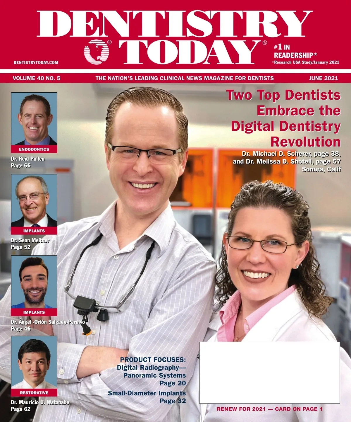
May 2021 · 76 pages
Dentistry Today — June 2021
Clinical articles, techniques, product news and continuing education for dentists. Read the full issue free in the digital edition.
Inside this issue
Excerpts from the issue. Open the digital edition to read any page in full.
- Page 8
Use Technology, But Don't Check Your Brain At The Door Despite all of the technology we see being used in many of our procedures, some require basic manual, visual, and mental skills. Single tooth restorations, oral hygiene, and diagnosis are all based on scenarios that also require our knowledge base. Much of this leads to conversations with our patients, which can easily be explained with basic show-and-tell; a written list of instructions; or even, heaven forbid, a simple even drawing. In our front-of-book columns, technology takes a back seat as Drs. Steven Kupferman and Jay Lee examine the perils of…
- Page 10
Early Detection Saves Lives! How often do we operate in the "run and gun" mode and omit the most important aspect of the patient visit—so much so that we may miss something right in front of our noses? We have been taught to look, listen, and feel lesions, like little TSA agents for oral cancer, but how much time are you reserving for that exam? Jumping out of the box of dentistry opens the door to save lives, one patient at a time, by treating the entire patient. Common causes of liability for failure to diagnose or delayed diagnosis of…
- Page 11
Jumping out of the box of dentistry opens the door to save lives, one patient at a time.... ple with inflammation post radiotherapy, the CS surgical mega soft (Curaprox) has more than 12,000 ultrafine filaments that are 6x co mm in diameter and guarantee perfect oral hygiene during special medical situations. More importantly, using an alcohol free mouthwash and a gentle toothpaste that does not contain sodium lauryl sulfate is a definite must with all your patients or your family. Using a toothpaste with natural enzyme activity can boost your saliva's natural defenses and help protect your patients from high…
- Page 12
Bone-Related Oral and Maxillofacial Conditions Steven B. Kupferman, DMD, MD, and Jay K. Lee, DDS, MD, discuss management, diagnosis, and treatment. Q: How prevalent are osteonecrosis and osteoporosis in dentistry? A: Osteoporosis is the most common bone disease in humans, affecting 1 in 2 women and 1 in 11 men. It can affect any bone in the body, including the jaw. However, this bone condition is not a part of normal aging. Certain drugs used to treat this condition can cause parts of the jawbone to become dead and exposed, known as osteonecrosis of the jaw. Osteonecrosis is rare; as…
- Page 16
Pulp Vitality Tester Helps Diagnose Patients' Issues EDGEWOOD, NY–Parkell's Digest 3 Pulp Vitality Tester electrically stimulates the tooth to test for vitality. Comparing the response to the stimulus of the suspected tooth with a known healthy tooth's response can help dentists hone in on the source of a patient's pain. A quarantine restrictions ease and patients return to the operatory to have long-overdue needs addressed, it has never been more important for clinicians to have every diagnostic tool at their disposal. With the ability to offer the additional diagnostic test of electric stimulation, dentists are discovering they can more easily…
- Page 17
TECHNOLOGY 17 Intraoral Scanner Produces Realistic Colors and Crisp Images SEOUL, South Korea–MEDIT's new i700 intraoral scanner is the company's next generation intra-oral scanner that builds upon the success of the award-winning i500. Powering the new unit is a super fast camera that captures up to 70 frames per second. This means more data with each pass, resulting in a smooth and quick scanning experience with realistic colors and a crisp image. The new camera and other internal components have been reduced in size, creating a new handpiece that is smaller and lighter, weighing just 2.3 oz (64 oz.). The…
- Page 18
TECHNOLOGY SPOTLIGHT 3D Printer Offers Features Rarely Seen in Economically Priced Models LOUISVILLE, Ky.–The VeriBuild LCD 3D Printer from Whip Mix offers features rarely seen in economically priced printers. Laboratories and dental practices can now print extremely precise models and accurate surgical guides with slice thicknesses between 25 and 100 µm. This open system printer is able to print 405-nm print resins using the proper file with its advanced software. Whether this is a person's first 3D printer or an add-on for increased production, he or she can count on consistently high print quality and high accuracy at an affordable…
- Page 20
Digital Radiography—Panoramic Systems Owandy Radiology The i-Max is the first wall-mounted pan system and is linked directly to one's network. All clinical and anatomical details can be viewed with maximum precision. The system is sleek and lightweight (136 lbs). It offers ALIS (Automatic Layers Integration System), which allows one to immediately and automatically select the best shots in order to display a perfect image. It includes an intuitive, user-friendly human-machine interface. The i-Max offers a superlative performance/investment ratio: lower purchase, delivery, and installation costs and easier maintenance. For more information, call (516) 858-5695, visit owandy.com, or email sales@owandy.us. FREEinfo, The…
- Page 22
Composite Solution's Elastic Properties Offer Easy, Failure-Free Placement THE VILLAGES, Fla–DMF Dental's Bright Temporary C&B is a new, self-curing composite for temporary crowns, bridges, inlays, and onlays. It is a unique combination of nanosized particles and ideally sized glass filler that provides enhanced composite strength. Its elastic properties provide easy, failure-free placement with the added advantage of the company's Chameleon Effect that matches shade, opacity, and fluorescence for a natural look. It offers 2 significant benefits, including (1) a short trim and finish time of 3 minutes, which eliminates the need for glazing and polishing, and (2) its convenience, which…
- Page 24
Composite Warming Dispenser Combines Several Innovations INDIAN LAND, SC-VisColor Dispenser is a composite capsule, rapid warming application device designed for VOCO's new, groundbreaking 2-in-1 thermo-viscous VisColor composite line. This sleek, ergonomically designed application device combines several innovations that enable the rapid warming of VisColor in 90 seconds while maximizing the working time to as much as 2 minutes, 35 seconds. The warmer uses innovative near infrared technology in tandem with VOCO's newly designed unit dose capsules. These easy access capsules are not only great for application but are also made from a special plastic that improves the heat transfer and…
- Page 26
Varnish Delivers Targeted and Sustained Release of Calcium, Fluoride SCHAUMBURG, Ill.- BISCO Dental Products' FluoroCal is a fluorideand calcium-releasing varnish that is indicated for application to enamel and dentin for the treatment of hypersensitive teeth. It delivers a targeted and sustained release of calcium and fluoride for more than 24 hours and provides immediate relief of sensitivity by penetrating and sealing dentin tubules. When tested, FluoroCal showed significantly higher fluoride uptake than other leading fluoride varnishes (data on file). Additionally, it contains Tri-Calcium Phosphate (TCP), which studies have shown may help strengthen enamel and build more acid resistant minerals when…
- Page 27
SPECIAL ADVERTISING SECTION 27 CASE STUDY A Clinician's Positive Results Using the Ortek-ECD A 22 year-old patient came to the office complaining of tooth sensitivity when eating sweets. Clinical examination showed obvious decay in the disto-occlusal pit on the maxillary first molar (Figure 1, site 3). Upon further examination, the mesio-occlusal pit (Figure 1, site 2) was suspicious, but there was no evidence of cavitation from visual and radiographic methods, making a treatment decision difficult. A second mesio-occlusal pit appeared sound (Figure 1, site 1). This case was ideal to evaluate the digital caries score indicated by the Ortek-ECD, as…
- Page 28
Industry News Scientific Metals Now Refining Dental Scrap Noting that metal prices are booming, Scientific Metals said that it is now refining dental scrap, including solids such as crowns, bridges, PFMs, PFIs, inlays, casting sprues, and buttons as well as grindings, floor and bench sweeps, amalgam, and filters. "Palladium, the silvery white metal, which is seen significantly in dental scrap, is nearing its all-time high. Dental scrap must be melted and assayed to accurately determine its palladium content, as palladium can easily be mistaken for non-precious metals with just a visual inspection," said company owner Dave Weinberg. The family-owned company…
- Page 30
Cancer... continued from page 28 detection by the immune system. The researchers aimed to better understand how this happens. When the immune system is functioning properly, the body's natural infection-fighting T cells help to identify and ward off carcinogenic cells, foreign viruses, and other invaders. But some cancer cells elude this immune response by means of protein molecules on their surfaces known as checkpoints that bind to similar molecules on the T cells, nullifying the immune cells' cancer-killing capabilities. Immunotherapy drugs called checkpoint inhibitors can be administered to help rectify this. These drugs turn off the cancer cells' checkpoint receptors,…
- Page 32
Small-Diameter Implants Shatkin F.I.R.S.T. Designed for single-tooth, multiple-tooth, and full-arch replacements (fixed and cemented roundhouses), Shatkin F.I.R.S.T. Mini Dental Implants provide the ability to replace and restore patients' teeth in one visit without the need to remove healthy tooth structure. Each implant offers ball and square engineering and is made of a strong titanium alloy that has an Osseam surface. The surface is impregnated into the implant, which allows for accelerated bone integration. For more information, call (888) 474-2854 or visit shatkinirst.com. FREEinfo, Nobel Biocare Ideal for areas with limited space, the Nobel-Active 3.0 is indicated for single-unit maxillary lateral…
- Page 34
continued from page 32 restore quality of life to your patients, often utilizing their existing dentures. The long-lasting TurLink silicone liner cushions and provides secure retention with easy insertion, making it ideal for patients with limited dexterity. For more information, call toll-free at (800) 323-3136 or visit dentatsusa.com. FREEinfo, Sterngold Sterngold is one of the leaders in the small-diameter implant market thanks to the MOR Mini Implant System. The popularity of MOR implants stems from their affordability, simplicity, versatility, and high potential to deliver patient satisfaction. Ranging from 2.1 to 3.0 mm, MOR Mini Implants are designed to help clinicians…
- Page 36
Reviewing Implant Technology and Material Innovations Beginning my implant career in 1984, I have seen many positive changes to the way we surgically place and prosthetically restore our implants. CBCT analysis and treatment planning has allowed the practitioner to understand vital anatomy, and with CAD/CAM design and milling technology used routinely, we are able to provide outstanding dental care to the most discriminating patients. Presented here are 3 outstanding teaching articles that will help our readers understand the most innovative treatment modalities available to all dentists. Early in my career, my mentor always promoted "visualizing the case finished before you…
- Page 38
Digital Clinical-Lab Workflows for Full-Arch Implantology INTRODUCTION Dentistry seems to be in the middle of an evolutionary tale, and possibly one of its revolution, about how one approaches the technical side of clinical practice. It seems that we, as clinicians, cannot escape the ever-present push of dentistry to incorporate digital techniques into our practices. One cannot read a newsletter, magazine, or journal or attend a trade show without the ever-present glitz and glamor of the latest in intraoral scanning technology, cone beam computerized tomography (CBCT), 3D printers, and milling machines. While it can be relatively straightforward to see some value…
- Page 40
Digital Clinical-Lab Workflows... continued from page 38 sufficient bone for implants. The surgical guide was 3D printed using a 3D printer (NextDent 5100 3D Systems) and UV cured to ensure biocompatibility. Working closely with a dental technician in the office, the intraoral scan optical files were imported into dental laboratory software (Dental System [s3shape]) for planning his maxillary and mandibular prostheses. Using photography and intraoral measurements, the teeth were positioned to plan for immediate dentures. Completed designs were imported into a milling machine software (Rs1VHP), the denture bases were milled using pink colored PMMA (Ivotion CAD [Ivoclar Vivadent]), and teeth…
- Page 42
Figure 10. A patient presents with reduced vertical dimension, failing dentition, and a request for dental implant treatment. Figure 11. Teeth were extracted, a surgical guide was adopted (3D Print Surgical Guide [SurtiRay]), and dental implants (Advanced Classic [Paltop]) were placed with the assistance of the guide. Figure 12. Healing abutments were removed, and contemporary fixed hybrid abutments (LOCATOR F-Tx [Zest Dental Solutions]) were placed. Figure 13. A panoramic radiograph (GreenCT) was made, confirming proper adaptation of abutments to dental implants. Figure 14. Final appearance of the maxillary overdenture abutments (LOCATOR R-Tx) and mandibular fixed hybrid abutments (LOCATOR F-Tx). Figure…
- Page 44
IMPLANTS ner (TRIOS) (Figure 15). The optical scan files were imported into dental laboratory planning software (Dental System), and maxillary and mandibular frameworks were designed for an overdenture and a fixed bridge. The STL file for the maxillary overdenture framework was sent to an outside dental laboratory for CAD/milled based 3D printing (Bertram Dental Lab, Menasha, Wis), and models were 3D printed using a desktop grade 3D printer (Form3B [Formlabs]) (Figure 16). The mandibular framework design file was imported into the in-house laboratory milling machine (Rs), and the framework was milled using a fiber reinforced polymer hybrid (TriLor [Harvest Dental])…
- Page 45
Patients with terminal and/or failing dentition are often seeking solutions to challenges that some clinicians may not address due to complexity, surgical concerns, and potentially patient derived factors. While complete arch reconstruction with dental implants can be a challenging diagnostic, surgical, and prosthetic modality, digital dental technology permits the surgeon and the restorative doctor to enhance prosthetic outcomes without enhancing the complexity of the procedures. Combining intraoral scanning, digital design, 3D printing, milling, and contemporary abutment systems permits the clinician to greatly simplify and create efficient work flows for the restoration of challenging cases. References: 1. Brown D. Oddities and…
- Page 48
Metal-Free, Implant-Supported... continued from page 46 t3: (c) root canal retreatment of tooth No. 15; (d) exodontia of the laminar implants of the second sextant (Figure 3) and placement of a removable mucous borne partial acrylic prosthesis to restore aesthetics and function immediately; and (e) after 5 months, planning the implant treatment. The CRCT scan showed a type IV to V alveolar ridge morphology, according to Atwood's1 classification (Figures 4 and 5). Antibiotic prophylaxis (amoxicillin 3 g one hour before surgery2) and rinsing with 0.15% chlorhexidine, in combination with 0.05% cetylpyridinium chloride, twice a day for 15 days, starting the…
- Page 50
Metal-Free, Implant-Supported... continued from page 48 around 3° between them should be chosen for optimal retention of the prosthesis. It is essential to keep this positioning key as it is the only way to be able to place the abutments in the same position. The copings (purple retentive or silver, non-retentive copings) were then placed on the abutments and checked to see that the gaps left in the frame of the BRWF did not interfere with the entry of the abutments and their copings. Once checked, a small amount of Vaseline was deposited inside the copings and left in place…
- Page 51
Figure 17. One-year follow-up OPG. BRWF is a material with optimal properties for the manufacturing of metal-free substructures. to the elastic deformation between both parts, an effect known as "cold welding."9 This union favors excellent bacterial seal, avoiding the bad odors typical of other implant systems when the abutments are removed. In such a system, the point surfaces must be dry as moisture can cause the abutment to come loose. This undesirable effect occurs more frequently in thick gingival biotypes or in those cases where the implant is positioned excessively subcrestally. The use of alcohol to clean the surface of…
- Page 52
Simplifying Implant Placement in the Maxillary Anterior INTRODUCTION Dental implant placement can be challenging based on the geometry of the bone present, especially in the maxillary anterior where the angle of the premaxilla requires a different trajectory then the prosthetic axis that will be placed during restoration. Traditional radiographs (periapical and panoramic) only provide 2D views of the 3D anatomy practitioners operate within. Thus, information regarding the inclination of the "bangle of bone" is absent, and implant placement requires a flapped approach to fully view the facial plate to aid in osteotomy preparation during implant placement. Even under these circumstances,…
- Page 54
Simplifying Implant Placement in... continued from page 52 tions if bone is adequate based on the desired implant position. A case demonstrating this technique and application with regard to a single implant placed in the anterior maxilla is presented here. CASE REPORT: SINGLE ANTERIOR IMPLANT PLACEMENT IN THE MAXILLA A 68-year-old male patient presented for consultation for a failing implant that replaced his maxillary left central incisor. Examination noted a facial 8 mm probable pocket and Figure 2. Components of the Guide Right system (DePhque): (a) Guide Posts, (b) Guide Sleeves, and (c) drills. Figure 3. A pilot hole was…
- Page 55
IMPLANTS the original trajectory (Figure 8, blue line). This new position allowed the implant to be placed buccally, centering it in the alveolar ridge based on the anatomy present as analyzed in the CBCT scan. A Guide Post with a 1.5 mm offset (Figure 2a, center) was inserted into the previously placed hole in the east wall in the offset position oriented toward the buccal. The offset Guide Post was rotated 0.5 mm mesially at 45° to reposition the upper extension of the offset post to a better prosthetic position (Figure 9). A 2-Piece Upper Removable Part (URP) (Figure 2a,…
- Page 56
Figure 17. The implant was placed level with the palatal crest. (a) an iGen 2-mm flat abutment (white part) was inserted in the graft. (b) The iGen titanium membrane was secured over graft material with a cover screw. Figure 18. The postoperative CBCT scan: (a) A cross section after graft augmentation shown with the 2 mm flat abutment and the iGen titanium membrane. (b) Tangential radiograph of the site following implant placement. perforated titanium membrane (A-i [MegaGen]) were tried in to verify the fit of the site and under the elevated flap (Figure 17;b). The titanium mesh membrane was secured…
- Page 57
Digital Workflows for Orthodontics and Clear Aligners INTRODUCTION The digital evolution in dentistry has had a profound effect on orthodontics, and particularly on the use and practice of clear aligners. While clear aligners were viewed for many years as a treatment only for adults with minor correction needs, they are now the treatment of choice of many clinicians for patients of all ages and treatment needs. The development of intraoral scanning and treatment planning software has led to an increase in the number of clear aligner cases and the increase of clear aligner treatment for comprehensive and complex cases. Innovation…
- Page 58
Figure 2. A full arch intraoral scan was taken to create a digital work-up and composite assessment of the patient's treatment needs (CS ScanFlow [Carestream Dental]). Figure 3. Digital models in articulation gave a visual representation of the patient's occlusion and demonstrated the need for clear aligner treatment to improve occlusion prior to restorative treatment (CS Model + [Carestream Dental]). Figure 4. Cone beam CT (Green CT [Vatech America]) was utilized for the comprehensive diagnosis and treatment planning of orthodontic treatment using micro-osseous perforations. ment. CBCT was first utilized in the United States at the Loma Linda University School of…
- Page 60
Figure 14. Use of the CS Model + software (Carestream Dental) to create an automated setup for clear aligner treatment. Figure 15. CS Model + software was utilized to create a sequence of clear aligners for comprehensive patient treatment. ers would be produced by the author with full control of potential movement challenges throughout treatment. The models created within the software were then 3D printed using a desktop-grade 3D printer (SprintRay Pro [SprintRay]) and the aligners (Zendura FLX [Bay Materials]) were fabricated and delivered to the patient the same day (Figure 15). CONCLUSION Orthodontic treatment with clear aligners has been…
- Page 62
Thermo-Viscous Bulk-Fill Composite Resin Restorations INTRODUCTION Restorations in the posterior are usually performed because of tooth decay, tooth fracture restoration damage, and filling replacement. They are probably one of the most performed procedures in restorative dentistry. Composite resin is the material of choice used in this direct approach because of the aesthetic results and longevity.¹ The incremental technique was largely used to execute posterior teeth restorations with regular composites. There are different approaches, such as horizontal layering, oblique layering, vertical layering, and stratified layering among others.² Regard-less of the technique used, the procedure can potentially lead to voids forming between…
- Page 64
Thermo-Viscous Bulk-Fill... continued from page 62 point inside the cavity and perform the extrusion, during which the temperature will drop even more, alleviating any concerns regarding pulpal health. Also, the dentin works as a temperature isolator, and we need to be aware of that fact when facing a very thin dentin thickness. Preheated composite resin induces a greater mobility of the monomers' molecules, increasing reactivity during photoactivation.⁵ Therefore, one could expect a higher degree of conversion when using a warmed composite resin to restore a tooth. Nevertheless, a significant increase in the degree of conversion is unlikely to occur when…
- Page 65
RESTORATIVE CASE 3 Figure 12. The old amalgams were removed from both posterior teeth while preserving the molar's marginal crest that had a tunnel preparation in the first clinical case. A composite resin was placed as material to alleviate any depth-of-cure concerns. Figure 13. First, the composite was used to seal the pulpal wall of the tunnel; then the material was filled to the proper level of the pulpal wall and light cured. Figure 14. The material capsule was warmed again for 3 minutes, and the cavity was then fully filled. Figure 15. The final result after finishing and polishing…
- Page 66
The Ultimate Root Canal Playbook: Part 1 I was a general dentist for 5 years. I know your root canal pain. I know that your endodontic education was limited, yet you are expected to be proficient at root canal treatment. I understand how difficult it is to perform root canals under poor conditions with limited materials, undertrained assistants, and minimal time. I also know that as a general dentist, I was terrible at performing root canal treatment and that I got sick and tired of feeling insecure and endodontically inadequate. Root canal treatment is a 3-quarter game—not a game of…
- Page 68
The Ultimate Root Canal Playbook... continued from page 66 deeper, but before the file encounters too much torque, disengage from the canal wall and back out approximately 3 mm. Repeat this process for 4 to 5 engagement/disengagements, and then pull out of the canal. Always shape with bleach in the canals. After each pass of shaping, irrigate with bleach and remove all of the accumulated debris. Keep in mind, we do not yet have working length but only an estimated working length from preoperative radiographs or CBCT scans. We are only flaring and cutting the middle part (middle flare). Negotiation…
- Page 69
Figure 6. WaveOne Gold Glider (Dentsply Sirona Endodontics). Figure 7. ProGlider (Dentsply Sirona Endodontics). So far, you have located the canals, performed a coronal negotiation approximately to to 15 mm down the canal with a #10 C 21 mm file, performed a coronal flare with a ProTaper Gold SX at 300 rpm, scouted the middle area of the canal with a #10 C 21 mm file, performed a mid dle flare with the ProTaper Gold S1 at 300 rpm, negotiated to patency and obtained an accurate working length. If the working length is 21 mm, then take a #10 K…
- Page 72
Systematic Approach to Prosthodontics Dinesh Sinha, BDS, PhD, interviews Jimmy Londono, DDS, director of the Ronald Goldstein Center for Esthetic & Implant Dentistry, about his prosthodontic protocol. Dinesh Sinha, BDS, PhD, Senior Technical and Marketing Manager, Dental Division, Kuraray America Jimmy Londono, DDS Q: Can you tell us about your practice? A: I am a prosthodontist and a clinical associate professor in the department of Oral Rehabilitation and director of the Ronald Goldstein Center for Esthetic & Implant Dentistry. As a prosthodontist, we evaluate different patients with different prosthetic needs every day. In some patients, the amount of breakage, wear,…