
July 2019 · 100 pages
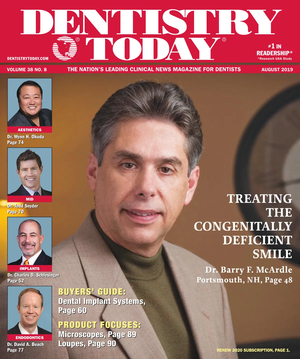
Dentistry Today — August 2019
Clinical articles, techniques, product news and continuing education for dentists. Read the full issue free in the digital edition.
Inside this issue
Excerpts from the issue. Open the digital edition to read any page in full.
- Page 6
The Implant Dentistry Option This issue of Dentistry Today focuses on implants. In 2015, we published our first Implants Today section, and it has been going strong ever since. Dr. Michael Tischler, our implant editor, and I make every effort to choose and place clinical articles into this section that will be of special interest to our readers—not only for those practitioners who already may have considerable experience in implant dentistry, but also for those who have not yet ramped up their participation in this growing arena. To lead things off this month, we have Dr. Barry McArdle presenting a…
- Page 8
The 90-Day Window for Trigeminal Nerve Injuries Trigeminal nerve injuries may occur as a result of many facial, oral surgical, and dental procedures despite the practitioner providing the highest level of care. Nerve injuries from third molar extractions, dental implant placement, endodontic therapies, orthognathic surgery, local anesthetic injections, or trauma are a known risk during many procedures, even when performed perfectly. Symptoms are wide ranging, but potentially devastating. Neurosensory deficits caused by dentoalveolar procedures can interfere with speaking, eating, drinking, smiling, and even intimacy. Without early intervention, these injuries may also develop into debilitating neuropathic syndromes. While most injuries are…
- Page 10
VIEWPOINT continued from page 8 nerve damage as soon as possible. Given the short window of time for optimal outcomes and the patient's lack of familiarity with nerve injuries and their effects, waiting for the patient to contact you regarding lingering changes in sensation or pain can result in a significant delay in diagnosing nerve damage. The patient might also disregard numbness for long enough that it becomes pain as a neuroma develops at the site of the injury. Symptoms to look out for may include: • Persistent loss or decrease of sensation in the lip, jaw, chin, cheek, or…
- Page 12
Consistent Restorative Excellence With Giomer Composites Jack D. Griffin, Jr., DMD Paramount in a busy clinical setting is the use of techniques and materials resulting in long-lasting restorations that promote oral health and are aesthetically pleasing. A versatile class of materials, featuring bioactive Giomer technology, meet all of these parameters with the bonuses of fluoride release and recharge and excellent handling. Beautifil II LS (Shofu Dental) is a packable Giomer composite with a unique filler content that delivers optimal surface hardness, compressive strength, wear, and aesthetics, making it a great choice for both posterior and anterior restorations. With the volumetric…
- Page 14
Team Morale and the Right Team Members Robert M. Pick, DDS, MS, discusses how having the right team members and amazing team morale is so important to achieving "Purple-Cow-Wow!" Q: When you lecture, you state that the team is always part of the "4 stages of a thriving practice," correct? A: Yes. I always state that there are 4 important basics that must occur for a practice to be outstanding: (1) do excellent dentistry, (2) have effective business systems, (3) have excellent relationship and communication skills, and (4) be a great team and have a great team environment. Without a…
- Page 16
You Must Believe Me Paul Feuerstein, DMD When an office uses digital chart notes, there are usually templates that are far more thorough than the paper charts, which had entries like "#5 MO composite." Preoperative image of a fractured molar. (Image courtesy of A.G. Khan, DMD, Billerica, Mass.) An image of a prep with a buildup. (Image courtesy of Dr. Khan.) With all the discussions on advanced dental treatment with new technology, there is an underlying issue that affects many of us. Some of us take insurance assignments, while others are fee-for-service and let the patients fend for themselves. I…
- Page 18
Treating Peri-implantitis Edward R. Kusek, DDS INTRODUCTION Treatment for peri-implantitis using traditional methods has been marginal at best. The dawning of laser dentistry has changed treatment from guarded to hopeful to successful. This article will present a case study that used numerous lasers to treat peri-implantitis. The First European Workshop defines peri-implantitis as inflammatory reactions in the hard and soft tissues, with loss of supporting bone, surrounding a dental implant exposed to the oral environment.¹ A successful dental implant is defined as immobile when tested clinically. Vertical bone loss is less than 0.3 mm annually following an implant's first year…
- Page 19
Use collagen membrane that is interwoven to prevent tissue invagination, and then place a PRF membrane over this collagen membrane before closing. The use of photo biomodulation aids in the healing process. Occlusal adjustments should be done at the time of surgery and 2 weeks after surgery. Consider the use of an occlusal guard. CASE REPORT Treatment of Peri-implantitis Using 9,300-nm CO2 and 2,780-nm Erbium Lasers This case showed early stages of bone loss and exudate present around the implant in position No. 9 (figure 1). The area is flapped with a CO2 laser (Solea [Convergent Dental]) with a setting…
- Page 20
Figure 1. Exudate present. Figure 2. Flap reflection was completed using a 9,300-nm CO2 laser. Figure 3. Surgical handpiece. Figure 4. Titanium brush being used at a slow speed to scrub the remaining remnants of granulation tissue around the implant. Figure 5. A 1,300-nm CO2 laser (Solea Convergent Dental) was used to detoxify the implant. Figure 6. BioCos bone/collagen material (Geistlich Pharma) with plasma from plasma-rich fibrin (PRF). Figure 7. Interwoven collagen membrane. Figure 8. PRF membrane. Figure 9. The tissue is epithelialized using an erbium laser. Figure 10. At 2 months postooperative. Figure 11. At 6 months postop (left)…
- Page 22
Spotlight Custom-Built Loupes Eliminate Eyestrain, Provide Ultimate Clarity LOMPOC, Calif.—DenMat's PeriOptix loupes are custom built to exact specifications, eliminating eyestrain and providing ultimate clarity, with a crisp and clear image across the entire viewing field. The loupes are available in 2.35x, 2.7x, 3.1x, and 4.0x magnifications for all optical needs. Frame options include a 2-position, adjustable nosepiece and 3-position adjustable arms to customize the declination angle, improve ergonomics, maximize comfort, and reduce fatigue. PeriOptix headlights offer powerful, lightweight, cordless and cordless options that mount directly to the frame to improve visual acuity while working in the oral cavity. PeriOptix has…
- Page 24
Spotlight System Provides Variety of Views To Fit Needs of Any Practice Laser Performs Procedures Without Injections, Leaving Patients Pain-Free ENGLEWOOD CLIFFS, NJ-The Litetouch All-Tissue Laser from integrated dental systems, in partnership with Light Instruments, offers one of the world's most versatile lasers in the United States. This game-changing laser, with built-in Laser-In-Handpiece technology, allows dentists to treat patients and do so more efficiently than ever before. This unique delivery system eliminates large fiber-optic lines and large articulating arms found on other lasers. Most LiteTouch procedures do not require injections, so patients are free from the pain and anxiety associated…
- Page 26
Spotlight System Combines Registration of Pressure Distribution of Occlusal Surfaces NASHUA, NH–Occlusense, a new system developed by Bausch, combines the traditional and digital registration of the pressure distribution of the occlusal surfaces. Occlusense handheld is a battery-powered device that uses a dual wireless network to transfer the recorded data to the Occlusense iPad app. The inductive charger provides a wireless power transfer. Fully charged batteries allow the usage of the device for up to 4 hours. The 6-μm Occlusense Electronic Pressure Sensor is used just like traditional occlusion test materials. The technologically advanced pressure sensors allow the recording of the…
- Page 27
The Dynamic Abutment Solution: Correcting Mal-Aligned Implants Tony Daher, DDS Most implant abutments and related components require parallelism within approximately 10° to function properly. Implant-angulation problems result from a non-parallel implant placement and in flared maxillary bone situations. This may preclude the use of a conventional implant abutment and corresponding attachment. When individual implants are not aligned or placed in the premaxilla, custom abutments may be used to correct for implant angulation variations, and the final implant restorations end up as cement-retained restorations. Our optimal restorative objective is to design screw-retained restorations, especially in complete-arch restorations. To avoid labial, buccal,…
- Page 28
Generational Advance in Antimicrobial Chemistry Points to Better Periodontal Treatment Outcomes Herb Moskowitz, DDS A recent breakthrough in iodine chemistry¹ permits the development of a new generation of antimicrobial agents that are significantly more effective and safer than commonly used povidone iodine (PVP-I) or chlorhexidine gluconate (CHX). These new iodine products have important applications in dentistry. They do not stain, they are not deeply colored, and they can be formulated in several pleasantly tasting flavors. Both CHX and PVP-I have established themselves as standards of care in infection control, globally.²⁻³ Each has withstood the test of time, having been proven…
- Page 30
Spotlight Matrix System's Ring Construction Stands Out SPRING LAKE, Mich.—Garrison Dental Solutions' newest innovation is the NiTin Sectional Matrix System. Used primarily for Class II posterior composite restorations, the system differentiates itself from the competition by its ring construction. Created from drawn-wire Nitinol, a product used in medical device applications including orthodontics, the material undergoes a process of molecular alignment that produces unparalleled spring qualities, ensuring separating forces that seem new even after hundreds of uses. Resiliency is reinforced with PEEK (polyether ether ketone), a super-plastic renowned for its durability, which is used exclusively in the construction of the backer…
- Page 32
Spotlight Light-Cured Desensitizer Provides Relief for up to 3 years in only 30 seconds ENCINITAS, Calif.–Shield Force Plus from Tokuyama Dental America is an ad-vanced, light-cured desensitizer that provides a pleasant, effective, and easy-to-use chairside solution for preventing post-restorative sensitivity and treating hypersensitive dentin. Its double block technology provides 2 layers of protection against sensitivity: The first layer penetrates the dentinal tubules for immediate relief while the second layer forms a protective barrier on the patient's dentin, protecting it from further abrasion and future sensitivity. When applied under a restoration, Shield Force Plus can increase bond strength, seal marginal areas…
- Page 36
Win a $250 amazon Gift Card! Take the Opioid Epidemic Survey amazon.com gift card $250 (only 5 questions) Enter for a chance to win online at survey Industry News DenMat Introduces Implant Services DenMat Holdings' lab services now include custom implant abutments and screw-retained crowns and bridges. These services enable custom restorative solutions for virtually any patient, simple or complex, from DenMat laboratory, according to the company. "The integrity of cases we receive are full arch restorations," said Randy Berry, DenMat director of laboratory operations. "We are seeing consistent growth in cases that include an implant as one of the…
- Page 37
Augmented Reality Lets Clinicians 'See' Their Patients' Pain Many patients, especially those who are syntactically or emotionally challenged, cannot communicate precisely about their pain. Now, researchers at the University of Michigan (U-M) have developed technology that helps clinicians "see" and map patient pain in real time through special augmented reality glasses. The technology was tested on two dental patients, and the researchers hope to one day include other types of pain and conditions. While it is years away from widespread use in a clinical setting, the feasibility study is a good first step for dental patients, said Alex DaSilva, DDS,…
- Page 38
Bio: Yasser Khaled, MD, is the son of a diplomat and has visited, worked, and studied in 15 different countries in continents around the world. He received a BDS degree and an MDSc degree in Oral Pathology from Ain Shams University in Egypt; an MMS degree in TMD/Orofacial Pain from the University of Alberta in Canada; and a GPR Certificate and an Oral Medicine Certificate from Atrium Health's Carolinas Medical Center in Charlotte, NC. He also received a Fellowship in Oral Surgery from Universiti Sains Malaysia. He has been awarded with hundreds of certificates from all around the world. He…
- Page 41
In First Impressions, George Freedman, DDS, gives readers a brief summary of products that have recently been introduced to dentistry, based on his clinical experience. He can be reached at (416) 473-0045 or via email at freedman@epdol.com. Omnichroma Tokuyama Dental America Ever since the first dental composite materials appeared in the 1970s, shade-matching restoration to the natural tooth or to ensure restorations has been a significant chairside challenge for the practitioner. Teeth come in a wide variety of natural shades, complicated by intrinsic staining, dietary staining, and aging. Moreover, most teeth are not monochromatic. They exhibit a variable color gradient…
- Page 42
CELLERANT BEST OF CLASS TECHNOLOGY AWARDS 2019 Earlier this year, the Cellerant committee met and voted on the Best of Class products of 2018 and a few that emerged in the first few months of 2019. There were several products that were previous winners and a few new innovations that have not been seen in dentistry before. The committee members talked with manufacturers, scoured dental meetings throughout the year, and sat through detailed presentations. Many of the products were evaluated in the members' practices, providing more opportunities to take a close look at the claims that were made and the…
- Page 45
CELLERANT BEST OF CLASS TECHNOLOGY AWARDS TechCentral OmniCore Henry Schein One | (833) 471-7253 | henryscheintech central.com/omnicore Most dental offices have a closet somewhere that contains the mysterious server. If you dare to open the door, usually there is a sign that says "do not touch." Someone in the office is usually dedicated to go in there: install software updates; check on backups, hoping that nothing catastrophic will occur; and call someone when there is an issue with a computer in the office. They are also are constantly worried about network problems, hackers, data breaches, and data backup. The OmniCore…
- Page 46
Implant Treatment Complications This month in Implants Today, we focus on complications related to dental implant treatment. Whenever a complex task is being performed, complications are often inevitable. The task of replacing a tooth or teeth as an implant dentist is a formidable task, as there are many variables involved for success. The mechanical challenges that we face are often compounded by the multiple biological factors presented by a patient. These biological factors include genetics, various medications, the patient's bite relationship, occlusion, and related functional habits; overall health and healing capacity; personal lifestyle such as smoking or alcohol consumption; and…
- Page 48
Treating the Congenitally Deficient Smile Barry F. McArdle, DMD Congenitally missing teeth are quite common in the adult population, being found on average in about 6.5% of individuals and with little to no change in those statistics over the past century.1 Furthermore, there is a strong association between having congenitally missing maxillary lateral incisor teeth and palatally impacted maxillary canine teeth.2 This article presents a case where a patient had just such conditions and how the author was able to achieve the patient's desired aesthetic outcome. CASE REPORT A 24-year-old female presented with dissatisfaction with her smile due to contour,…
- Page 50
IMPLANTS FIGURE 2. The patient's initial clinical presentation with the same conditions. FIGURE 3. The patient one week after placement of implants at the upper canine positions and insertion of their provisional crowns. Note the convexities present at the incisal aspects of the central incisors. FIGURE 4. The patient's pre-impression presentation with maxillary canine teeth at the lateral incisor positions after preparation for the lateral-incisor-shaped provisional restorations. Note the fuller incisal aspects of the central incisor teeth. FIGURE 5. The final impression of the canine implant abutments and the prepared canines occupying the lateral incisor positions. FIGURE 6. The patient…
- Page 52
Guided Surgery: Accurate, Predictable, and Economical Charles D. Schlesinger, DDS Figure 1. Differences between a conventional CT scanner and a CBCT scanner. Figure 2. The 3 types of surgical guides. INTRODUCTION As technology advances, patient treatments advance too. The advent and continual development of cone beam computed tomography (CBCT) machines and guided surgery continue to provide clinicians with incredible tools to optimize patient care. The technique used in CBCT has been applied in medical imaging since 1982.¹ The first CBCT units were developed and sold in Europe in 1996 by NewTom. These units were very large and expensive. As a…
- Page 54
Figure 3. A bone reduction guide for an uneven ridge. Figure 4. Implant delivery mount. Figure 5. A surgical guide key. CBCT Guided Surgery: Accurate... continued from page 52 sue reflection necessary.5 One other style of guide is a bone-reduction guide. This type of guide is used similarly to a tooth-reduction guide that your lab may send you when a preparation is underprepped for a crown. It is used in the same manner as a bone-supported guide. Once placed, the portion of the ridge that is sticking up beyond the guide is reduced (Figure 3). This form of guide is…
- Page 56
IMPLANTS means you lose some of the benefits that likely encouraged you to buy the specific implant you are using. Guided Surgery Only vs Guided Surgery and Guided Placement Guided-surgery-only kits will guide your osteotomy drills to the appropriate position, depth, and angulation. Then the guide is usually removed, and the seating of the implant in the osteotomy is done by hand. What does this mean for you? It means that the final position of the implant may be slightly off from what was planned. This discrepancy can increase, especially if you are placing in an area of the mouth…
- Page 58
Figure 25. Pre-op radiograph of lower left quadrant. Figure 26. Intraoral view. Figure 27. Guided treatment plan. Figure 28. Try-in of guide. Figure 29. Alignment drill. Figure 30. Pilot drill. Figure 31. The pilot drill was taken to full length. Figure 32. Final 5-mm-diameter osteotomy former. Figure 33. Removing the mounted implant from the carrier. Figure 34. Both implants, fully seated through the guide. Figure 35. Placement of 3-mm healing abutments on both implants. Figure 36. Final radiograph, verifying position of implant Nos. 18 and 19. CBCT Guided Surgery: Accurate... continued from page 56 tance from the top of the…
- Page 59
and tightened with a driver. This mounted implant was then delivered (Figure 19) through the guide and advanced to its final position with a ratchet wrench (Figure 19). The final seating position is again determined when the stop on the fixture mount comes into contact with the sleeve in the guide (Figure 20), and the flat on the mount matched the flat on the sleeve (both are hexagonal in shape). The mount was then removed and the guide was removed from the patient's mouth (Figure 21). Next, an ISO reading was taken using an Osstel (Osstel) (Figure 22). With the…
- Page 60
Dental Implant Systems Bicon Dental Implants The SHOREÅ implant design comprises plateaus, sloping shoulders, and a bacterially sealed, 1.5-locking taper implant-to-abutment connection. With the plateau design, cortical-like bone forms around and between each plateau, which allows for the routine use of 5.0 mm short implants. The sloping shoulder provides the necessary room for bone to support interdental papillae that is gingivally aesthetic. Bicon's 360° universal abutment positioning provides for the revolutionary cementless and screwless integrated Abutment Crown, consistently offering a nonmetallic aesthetic gingival margin. For more information, call (800) 882-4266. FREEinfo, BioHorizons Tapered Implants have been developed based on more…
- Page 62
continued from page 60 HIOSSEN ETiii dental implants are manufactured with pure, medical-grade titanium metal and sculpted with a sandblasted and acid-etched (SA) surface. The ETiii SA implant shortens bone healing time, while providing convenient placement and initial stability because of its optimal body design. Last March, the ETiii NR implant was released. It has a nanolayer of bioresorbable apatite coating with hydrophilic properties. In addition, the new ETiii SA with a 2.3-mm diameter is a conventional implant for restrictive spaces of the aesthetic zone. For more information, call (888) 678-6001 or visit the website hiossen.com. FREEinfo, Implant Direct The…
- Page 64
DENTAL IMPLANT SYSTEMS continued from page 62 minimally invasive piezo-surgical placement and conical taper leading to thread connection, the implant is designed and tested for long-term stabilization for fixed or removable crown and bridge applications. All implantologists will be interested in this implant. For more information, call (800) 243-7372 or visit the website parkdentalresearch.com. FREEinfo, PerioSeal The PerioSeal Implant System provides today's general dentist and specialists the simplest yet most advanced system marketed in the United States for addressing the issues of highest concern to patients and dentists. Its unique design incorporates safety features, lower costs, simple crown-and-bridge dentistry, and…
- Page 66
DENTAL IMPLANT SYSTEMS continued from page 64 and applicable for a wide range of indications. The company's primary goal is to make a guided surgery protocol easy and simple to practice. It offers a fully guided Pylon kit and the utilization of CAD/CAM expertise allows IBIS Implants to design and manufacture a patient-specific prosthesis prior to surgery with excellent predictability. For more information, call (866) 435-8247 or visit the website urisimplants.com. the confidence in the Prettau zirconia. With custom shades, custom shapes, no chipping, no staining, to acrylic, and less prosthetic space needed than a hybrid bridge, the Prettau zirconia…
- Page 70
Patient-Driven Aesthetics A Conservative Approach Todd Snyder, DDS INTRODUCTION Dentists often want a routine or list to guide us in our approach to everything we do in our practices. We want our jobs systematized to the point that we have developed principles and philosophies such as Golden Proportionality, Golden Percentage, ideal length-to-width ratios, and more that allow us to define various aspects of a pleasing smile. These are all important concepts for a dentist to understand and study. However, what if patients want something different—something that falls outside of the parameters and standards that we have been trained to follow?…
- Page 72
Figure 4. Baseline acrylic mockup in place, allowing the patient to visualize the new smile design. Figure 5. Retracted view of the potential new teeth appearance with the baseline acrylic mockup. Figure 6. Depth cuts were placed into the acrylic mockup to guide the minimal bone preparation. Figure 7. Reduction of the acrylic and tooth structure between each depth cut. Figure 8. All of the facial surfaces shown here had been reduced and combined. Figure 9. A photo of the shade tab was sent to the lab team for color evaluation. Figure 10. The final impression, using a customized tray…
- Page 73
MINIMALLY INVASIVE DENTISTRY 73 procedure because she could see when she smiled that the gumline was not a concern for her cosmetic appearance. This ability to visualize the potential case outcomes so realistically provides instant insight and helps educate decisions as to how the treatment will proceed. After approval of the cosmetic appearance is obtained, the patient can either return another time or have the procedure done the same day. Clinical Protocol The next step is to place depth cuts into the acrylic (Figures 6 and 7). If the initial appointment was just mock up the case, then the patient…
- Page 74
Correction of Extremely Dark Anterior Teeth Wynn H. Okuda, DMD INTRODUCTION Anterior tooth discoloration continues to be an aesthetic concern and a major source of embarrassment for patients. The problem can either be of extrinsic or intrinsic origin, so the clinician must determine this prior to treatment. Extrinsic stains can be removed through oral hygiene and bleaching treatment, as they are stains that build up on the surface of the dentition over time. However, intrinsic stains are internal stains that must be treated through methods that depend on the origin of etiology. Depending upon the type and/or cause of the…
- Page 76
Figure 2. Dentin adhesive resin placed prior to masquing the dark stain with opacious dentin composite resin. Figure 3. After the bonding of the spacious dentin shade, the porcelain veneer preparations were finalized. Figure 4. The final result was more aesthetic because a non-opaque veneer was used to correct this problem. to bond in the porcelain veneers to achieve a bright overall effect. Final bonding was accomplished by light curing (Sapphire Plasma Arc Curing Light[DenMat]) to the porcelain veneers along the facial and lingual surfaces. The final finish of the restorations was accomplished with a series of finishing diamond burs…
- Page 77
Apical Lesions: Diagnostic Considerations David A. Beach, DMD, MS INTRODUCTION "My dentist saw a dark spot on the x-ray." This statement is commonly heard from patients in endodontic offices on a routine basis. What the patients are referring to is, of course, a possible abscess or some other form of periradicular radiolucency (PRRL). The diagnostic question the practitioner must answer is "What treatment is necessary?" Do all "dark spots" need treatment? The following case reports will highlight some of the diagnostic possibilities for various PRRLs. CASE 1 The True Dental Abscess One of the radiographic manifestations of a necrotic tooth…
And 15 more pages — read the whole issue.